The magnetic resonance force microscope
(MRFM) is a novel scanned probe instrument which combines the three-dimensional
imaging capabilities of magnetic resonance imaging with the high sensitivity
and resolution of atomic force microscopy. It will enable non-destructive,
chemical-specific, high-resolution microscopic studies and imaging of subsurface
properties of a broad range of materials. This technology holds clear potential
for atomic-scale resolution.
New magnetic materials and devices with unprecedented
capabilities and levels of performance are now being created by tailoring
the structure and composition of multi-component materials at the nanometer
scale. The buried interfaces between the various components of these new
materials play a central role in determining their functional behavior.
We are presently applying the emerging capabilities of the MRFM to the
study of layered magnetoelectronic materials. The MRFM will enable very
high resolution studies of the structural and magnetic properties of the
buried interfaces.
|
| High
Resolution Force Detected Magnetic Resonance Imaging |
Force Detection of
Magnetic Resonance
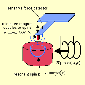
The magnetic resonance force microscope (MRFM) is a microscopic imaging
instrument that mechanically detects magnetic resonance signals by sensitively
measuring the force F=m·ÑB
between
a permanent magnet that provides ÑB,
and the spin magnetization m. Periodically modulating this force
by modulating m alters the oscillation amplitude of a high Q, low
spring-constant micro-mechanical resonator (cantilever or bridge) such
as is used presently in AFM.
High Resolution Magnetic
Resonance Imaging
The magnetic field gradient of the small magnetic tip serves a second key
role: it defines the spins (electronic or nuclear) which will be probed
in the magnetic resonance experiment. The resonance frequency of a spin
is proportional to the magnetic field it experiences:
w=gB(r),
only the resonant spins are effected by the magnetic resonance experiment.
As in conventional magnetic resonance imaging (MRI) the spatial variation
of B allows us to probe only those spins whose resonance frequency matches
the frequency wo of the applied rf
field, that is those spins residing in the "sensitive slice." |
|
Magnetic
Multilayer Materials
|
High sensitivity
magnetic field detectors for high density magnetic information storage
devices.
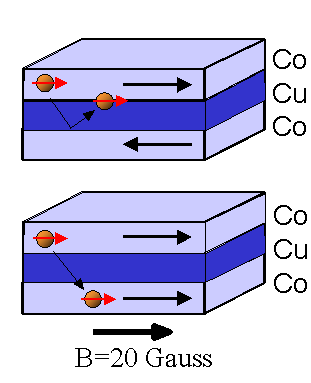
The field of magnetoelectronics is experiencing a huge resurgence of interest
and a new influx of research effort. The driving force is the technological
quest of the $100B/yr magnetic recording industry for increased storage
density in recording media. Further impetus also comes from the semiconductor
industry, in the area of memory: ideal non-volatile memory elements are
sought that can be produced, and addressed, in massive arrays. The
growth of high quality magnetic multilayer materials is providing the foundation
for a range of important modern advances in both fields, and offers
a new range of possibilities. The quality of such materials, upon which
the performance of these devices so strongly depends, is affected not only
by the purity of the materials grown, but, crucially, by the properties
of the interfaces between different layers. For example,
in magnetic multilayer materials displaying "giant'' magnetoresistance
(GMR), device performance is critically dependent on the microscopic characteristics
of the buried interfaces. GMR-based read heads have enabled up to
a 50% increase in magnetic storage density and have thus become a central
new technology for the industry. A second example is the recently developed
spin injection devices, which offer a new approach to non-volatile magnetic
memory. Although the physics of these new types of magnetic
systems is highly dependent upon interfacial characteristics, remarkably
little is known about the microscopic morphology of these interfaces.
Nor are the microscopic mechanisms that determine interfacial magnetic
properties well understood. MRFM studies will enable unprecedented
understanding of structural and magnetic properties of buried interfaces
and microstructure; this will enable development of materials with improved
properties. |
 |
Fully Scanning Cryogenic MRFM with Detector Mounted Micromagnetic Probe
A general purpose MRFM must have the microscopic ferromagnetic probe tip mounted
on the detector that can be scanned over arbitrary samples. This
presents signal detection difficulties because the ferromagnet couples
to the time varying fields that are necessary to excite magnetic resonance.
This leads to forces on the mechanical detector that can greatly exceed the
forces generated by magnetic resonance.
Nonetheless, this geometry is essential for a general purpose scanning
microscope.
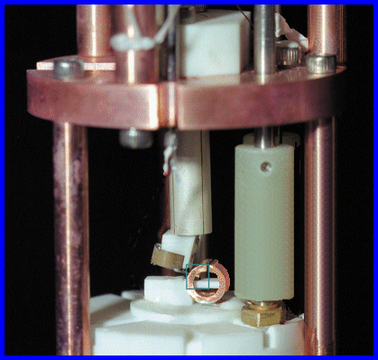
We have constructed the cryogenic MRFM shown here. It operates
in vacuum at liquid He temperature. The piezoelectric scan tube visible in
the center of this photograph is about one inch in length and will
allow scanning over a region 16 microns in diameter at low temperature.
The cantilever with a micromagnet mounted on it is mounted on the angled
block at the end of the tube.
The sample mount is located inside the cylindrical copper NMR coil.
For electron spin resonance and Ferromagnetic resonance, this coil is
replaced by a microwave micro-strip resonator. The present sensitivity
of this microscope is approximately 10
mB.
|
 |
Collaborators
|


